Combined SICM/SCEM
We have performed pilot experiments to design a special probe tip for simultaneous scanning electrochemical/ion conductance microscopy, which consists of a micropipette for ion current measurements, surrounded by a gold or platinum ring electrode, and an outermost electrophoretic insulating sheath. The gold or platinum was sputtered to produce 100-200 nm coating on the glass micropipette and then pipette was electrophoreticly coated with insulating paint. Then the very tip was cut by ion beam to expose a gold or platinum ring. This micropipette surrounded by a gold or platinum and an insulating layer of electrophoretic paint was filled with electrolyte and attached to a z piezo of SICM and has been employed as the feedback signal to regulate the tip-substrate distance. This probe could serve as a SECM microelectrode and allow simultaneous accurate topographic measurements, along with SECM imaging (Fig. 1). Ring electrodes can be fabricated from different metals or carbon since the electrode material is known to be important to detect different chemical species. Recently the fabrication of a similar probe based on a nanopipette was demonstrated

Figure 1 (A) Schematic illustration of SECM/SICM measurement. (B) SEM micrograph of SECM/SICM aperture. (C) Cyclic voltammogram of a SECM ring electrode in a 0.5 mM FcMetOH in 0.1 M KCl solution.
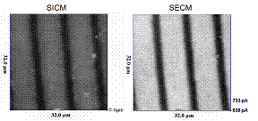
The feasibility of this approach has already been confirmed by obtaining simultaneously topographic and electrochemical images of an interdigitated platinum electrode (Fig. 2) and live cell imaging (Fig. 5) with a resolution of about 500 nm, limited by the size of the probe. However further work is now required to reduce the probe size and optimise its performance.

Figure 2 . Faradiac (right) and topographic image (left) using ion current feedback signal of Pt band micro-array. The electrodes held +0.3 vs. Ag/AgCl and +0.5 vs. Ag/AgCl, respectively.
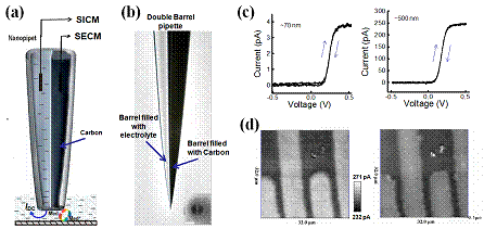
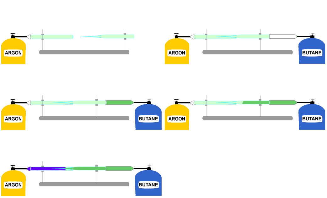
We have performed pilot experiments to design another type of combined SICM/SECM probe. Here the micropipette is fabricated with a double barrel configuration; one barrel being the microelectrode for SECM, and the other being used to contain an electrolyte for SICM. The double barrel pipette can be pulled from a “theta glass” capillary (whose cross-section resembles the Greek capital letter Theta, in which a straight septum divides the circular cross section into two isolated compartments (Fig. 3). We have used these pipettes previously for SICM and controlled deposition. For SICM/SECM one barrel is coated internally with the conductive carbon, by pyrolytic decomposition of butane. After deposition, electrical contact is established with the internal conductive layer by inserting a conductive wire through the top end of the pipette barrel and into contact with tip region. If the resulting conductive layer inside the barrel is sufficiently thick to fill and block the tip of that barrel, it provides an effective SECM microelectrode, where the exposed end of the conductive carbon is surrounded by the insulating barrel wall. The second barrel is unmodified, and filled with electrolyte and is used for SICM control. The images in Figure 4d show the feasibility of this approach. The resolution was about 500 nm in these images showing the feasibility of this approach but again further work is required to reduce the probe size for higher  resolution imaging.
Figure 3. (a) Schematic illustration of SECM/SICM measurement. (b) Optical image of double barrel pipette with one barrel filled with pyrolitic carbon. Insert is a SEM micrograph of the pipette opening. (c) cyclic voltammogram (50 mV/s) of ~70nm (left trace) and ~500 nm (right trace) SECM carbon electrodes in a 2.5 mM FcMetOH in 0.1 M KCl solution. (d) Faradiac (left trace) and topographic image ((right trace) using SICM current feedback signal of Pt band micro-array. The electrodes held +0.2 vs. Ag/AgCl and +0.5 vs. Ag/AgCl respectively .

Figure 4 Intagration carbon electrode to the nanopipette by pyrolytic decomposition of butane
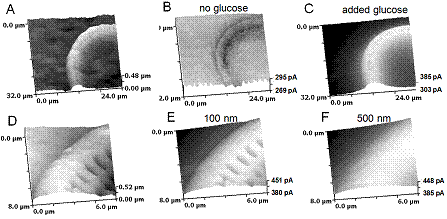
The images in Figure 5 show the feasibility of recording enzymatic activity with submicron resolution but again further work is required to reduce the probe size for higher resolution imaging. The figure 5E and F highlights the necessity of the precise control of the distance between the electrode and the sample.

Figure 5 Topography (A & D) and faradiac (B,C,E &F) images of the glucose oxidase (GOx) immobilized substrate. D –F images were obtained simultaneously where images E and F were recorded at 100nm and and 500nm from the surface respectively. The 400nm SECM /SICM Au ring electrode was held +0.5 vs. Ag/AgCl (for SECM) and +0.2 vs. Ag/AgCl (for SICM) and was used for imaging in PBS solution.
